Creating places that enhance the human experience.
| Page 3 |
31e. Technical Notes and Review1e-1. Homogenization mediaThe homogenization medium (HM) often has been specifically tailored to fractionation ofDrosophila membranes. In the example given solutions are buffered with Tris but Hepes, Tricine ortriethanolamine (at the same concentration) may be used if preferred. It is unlikely that the type ofbuffer significantly influences the homogenization or fractionation, although with mammalian cellstriethanolamine does seem to offer some particular advantages in homogenization efficiency.The preparation of a Working Solution (Solution I) as described, ensures that the concentrations ofKCl, EDTA and Tris buffer are constant throughout the gradient, while the sucrose and iodixanol act asosmotic balancers to maintain an approx. constant osmolality. If this is deemed unimportant thegradient solutions may be prepared directly from OptiPrep™, but if this option is chosen then theconcentrations of KCl, EDTA and buffer will decrease with increasing solution density.Beronja et al [1] adjusted the KCl concentration in the homogenate (50 mM) as described in theprotocol, but Papoulas et al [3] adjusted it to 100 mM. If the KCl concentration is adjusted to 100 mMKCl, then all subsequent solutions used (Solutions E-H) should be similarly adjusted.Tan et al [4] used 0.25 M sucrose, 1 mM EDTA, 1 mM DTT, 10 mM HEPES, pH 7.4 as anhomogenization medium in their proteomic studies and in some cases the osmotic balancer is NaCl andnot sucrose, e.g. 150 mM NaCl, 0.2 mM EGTA, 100 mM Tris, pH 7.4 [5]1e-2. Ultracentrifuge rotorsChoose whichever swinging-bucket rotor is most suitable for the amount of material available (seeStep 6). The iodixanol gradient centrifugation is carried out in a near-vertical rotor. The gradient isformed partly by self-generation, partly by diffusion. Other rotors with different sedimentation pathlengths may be suitable but the optimal centrifugation conditions will require investigation. Near-vertical rotors are preferred over vertical ones because any very dense particles will form a welldefined pellet close to the bottom of the tube (as in a fixed-angle rotor), while in a vertical rotor anydense material will pellet along the entire length of the wall of the tube and may contaminate fractionsduring unloading. The use of Beckman Optiseal™ tubes is recommended because of the ease of useand the ability to use a variety of options for gradient unloading (see Step 14); for other tubes such asheat-sealed tubes, tube puncture is the only safe and reliable option. For more information self-generated gradients see Application Sheet S04.1e-3. Preliminary purificationFor the analysis of the major membrane fractions (plasma membrane, Golgi and ER) the 3000 gsupernatant may be simply centrifuged at 100,000 g to pellet the microsomes [3] or partially purifiedby sedimentation through a sucrose cushion [1]. The latter will more effectively remove solubleproteins, which will remain principally in the sample zone. The 100,000 g step was replaced byPapoulas et al [3] by a 20,000 g step if the aim was to isolate primarily the Golgi for use in vitroincubations.1e-4. Gradient variationsAdolfson et al [6] adopted a simpler iodixanol gradient format; the post-nuclear extract wasadjusted to 26% (w/v) iodixanol to form a self-generated gradient at 300,000 gav in a near-verticalrotor, while Niimura et al [7] used a discontinuous 2.5-30% (w/v) iodixanol gradient.1e-5 Gradient layeringAlthough underlayering with a syringe and metal cannula is the recommended method for makingdiscontinuous gradients, overlayering maybe more convenient since the tubes need to be filled exactlyto the bottom of the neck. For more information on gradient construction see Application Sheet S03. Ifnecessary, adjust all volumes proportionately so that tubes (after sample application) are properly filledaccording to the manufacturer’s instructions.
| Page 4 |
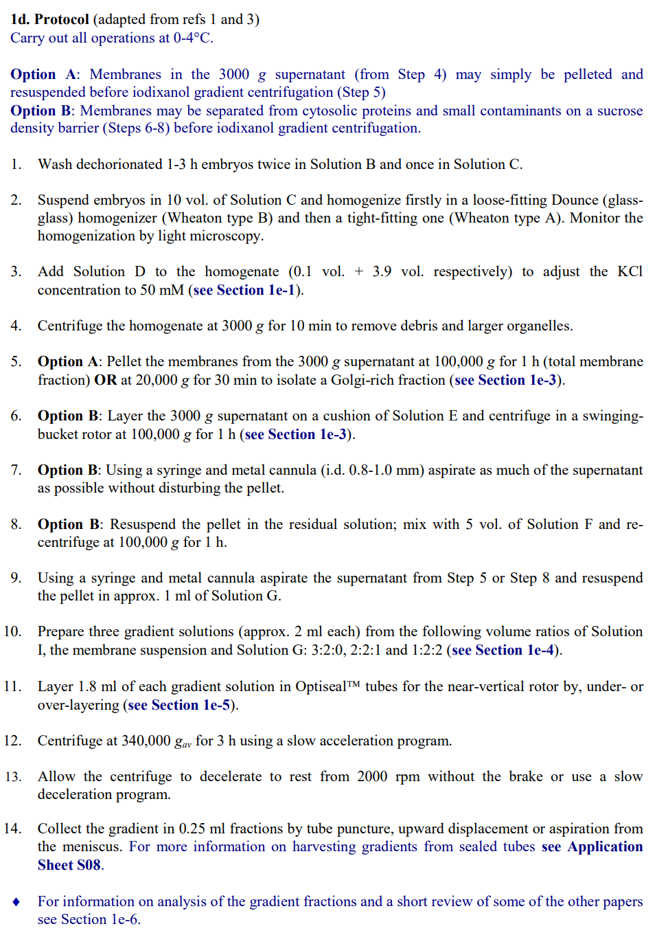
41e-6. Method reviewIn the gradient system described in thisOptiPrep™ Application Sheet, the Golgi, ERand PM banded approximately as shown inFigure 1. The Golgi was identified by theLava lamp protein, the ER by BiP and the PMby α-spectrin, which was also present in theGolgi region [3]. Unlike most mammaliancells the Golgi was denser than the ER; butlike mammalian cells, the PM banded close to the top of the gradient. Dynein also co-banded with theGolgi but was also present in some denser fractions. These gradients were used in characterization ofthe Sec6 component of the exocyst complex [1], the dynein based motility of Golgi membranes [3] andto confirm the association of dLgl and dFmr1 with the Golgi [8]. The method has also been used inproteomic studies [9] and to identify an ER location for the Seele protein [10].In the 2.5-30% discontinuous iodixanol gradients used by Niimura et al [7] the ER banded at ahigher density than the Golgi. In the self-generated gradients reported by Adolfsen et al [6], synapticvesicles banded close to the top of the gradient and clearly discriminated low-density vesiclescontaining Synaptotagmin 1 from denser ones containing Synaptotagmins 4 and 7. Paneels et al [11]used a 5%, 30%, 40% iodixanol flotation gradient to band the plasma membrane at the 5%/30%interface (see Section B).A 10-40% (w/v) iodixanol gradient (250,000 g, for 3 h) separates the plasma membrane from thecytoskeleton [12]. A simple three-layer gradient in which a 1000 g supernatant in 40% iodixanol islayered beneath layers of 5% and 30% iodixanol [5,13] centrifuged at 100,000 g for 3 h results in theplasma membrane banding below the 5% layer. Tan et al [4] underlaid a 2000 g/5 min supernatant with6% and 8% (w/v) iodixanol and centrifuged at 100,000 g for 90 min to concentrate the membranes atthe interface of the two iodixanol solutions. The recovered membranes were adjusted to 12.5%iodixanol and centrifuged in a Beckman VTi65.1 vertical rotor for 1 h. The method resolved PM,Golgi, ER and mitochondria.♦ For some more recent publications see Section 42. Isolation of lipid rafts (as detergent-resistant membranes)2a. BackgroundRietveld et al [14] were the first to report isolation of lipid rafts from Drosophila using flotation iniodixanol gradients. A crude PM fraction was first produced from a post-nuclear supernatant of thehomogenate (adjusted to 1.4 M sucrose) by flotation through a layer of 1.22 M sucrose. Hoehne et al[15] used a similar approach. Paneels et al [11] and Eroglu et al [16] adapted this plasma membraneisolation method to iodixanol. The crude low-density membrane fraction was then extracted withTriton X-100 and the lipid rafts isolated by flotation through a discontinuous iodixanol gradient. Zhaiet al [17] prepared lipid rafts from both Drosophila and from the Drosophila S2 cell line directly froma 5000g supernatant of the homogenate, without a preliminary preparation of a crude PM fraction.♦ The method below is adapted from refs 11, 14 and 16. Some of the variants are described in theTechnical Notes and Review Section (Section 2e).2b. Solutions required (see Box on next page and Section 2e-1)A. OptiPrep™B. Wash Solution 1: 0.9% (w/v) NaCl, 0.1% (w/v) Triton X-100C. Wash Solution 2: 0.9% (w/v) NaClD. Homogenization Medium (HM): 0.3 M sucrose, 150 mM NaCl, 0.2 mM EGTA, 100 mM Tris-HCl,pH 7.5E. TNE: 150 mM NaCl, 0.2 mM EGTA, 100 mM Tris-HCl, pH 7.5High densityLow densityGolgiERPlasma membraneFigure 1. Distribution of Drosophila membranes in iodixanolgradient; for more information see text. Data adapted from ref 3
| Page 5 |
5F. Optiprep™ Dilution Buffer: 150 mM NaCl, 1.2mM EGTA, 100 mM Tris-HCl, pH 7.5G. Optiprep™ Working Solution (50% iodixanol):Dilute 5 vol. OptiPrep™ with 1 vol. of Solution F2c. Ultracentrifuge rotor requirements (seeSection 2e-2)Swinging-bucket rotors: approx. 27 ml tubes (e.g.Beckman SW28) and approx. 5 ml tubes (e.g.Beckman SW55)2d. ProtocolCarry out all operations, except step 1, at 0-4°C.1. Wash the dechorionated embryos twice inSolution B, three times in Solution C.2. Wash the embryos twice in Solution D.3. Suspend washed embryos in 10 vol. of Solution D and homogenize firstly in a loose-fitting Dounce(glass-glass) homogenizer (Wheaton type B) and then a tight-fitting one (Wheaton type A).Monitor the homogenization by light microscopy (see Section 2e-3).4. Centrifuge the homogenate at approx. 3000 g for 10 min to remove nuclei and debris.5. Adjust the iodixanol concentration of the 3000 g supernatant to 40% (w/v) iodixanol by mixing 1vol. with 4 vol. of Solution G (see Section 2e-4).6. Prepare solutions of 30% and 5% (w/v) iodixanol from Solution G and Solution D (volume ratiosof 3:2 and 1:9 respectively).7. Distribute the 3000 g supernatant (in 40% iodixanol) equally amongst tubes for the 27 mlswinging-bucket rotor and layer 10 ml and 5 ml respectively of the 30% and 5% iodixanolsolutions on top to fill the tube (see Section 2e-5).8. Centrifuge at 100,000 g for 3 h.9. Collect the plasma membrane enriched fraction from the 5%/30% iodixanol interface and dilutewith 3 vol. of Solution E.10. Pellet the membranes at 50,000 g for 30 min.11. Aspirate the supernatant; resuspend the pellet in Solution E and repeat Step 10.12. Resuspend the pellet in 0.5 ml of Solution E and mix with an equal volume of Solution Econtaining 2% (w/v) Triton X-100 (or other chosen detergent at twice the required concentration).13. Keep at 4°C for 30 min to solubilize the detergent-sensitive membranes.14. During the solubilization prepare solutions of 21%, 15% and 6%(w/v) iodixanol by dilutingSolution G with Solution E at volume ratios of 2.1:2.9, 1.5:3.5 and 0.6:4.4 respectively. Note thatthere are important published variations in the density of the gradient solutions (see Section 2e-6).15. Mix the suspension from Step 13 with an equal volume of Solution G.Keep the following stock solutions at 4°C:1 M Tris (free base): 12.1 g per 100 ml water1 M NaCl: 5.84 g per 100 ml water100 mM EGTA (free acid): 3.80 g per 100 ml water (pH11-12)Solution D: Dissolve 20.5 g sucrose in 100 ml water;add 30 ml, 20 ml and 0.4 ml respectively of NaCl, Trisand EGTA stock solutions; adjust to pH 7.5 with HCland make up to 200 ml.Solution E: To 100 ml of water add 30 ml, 20 ml and 0.4ml respectively of NaCl, Tris and EGTA stock solutions;adjust to pH 7.5 with HCl and make up to 200 ml.Solution F: To 50 ml water add 15 ml, 10 ml and 1.2 mlrespectively of NaCl, Tris and EGTA stock solutions;adjust to pH 7.5 with HCl and make up to 100 ml.
| Page 6 |
616. In tubes for the approx. 5 ml swinging-bucket rotor, over layer 2 ml of the sample with 1 ml eachof the 21%, 15% and 5% (w/v) iodixanol solutions (see Section 2e-5).17. Centrifuge at approx 150,000 gav for 5-6 h (see Section 2e-7).18. Allow the rotor to decelerate without the brake below 2000 rpm or use a controlled decelerationprogram.19. The lipid rafts band as a visible layer at the top interface. Harvest this layer or collect the gradientin 0.25-0.5 ml fractions by tube puncture, upward displacement or aspiration from the meniscus.For more information on harvesting gradients see Application Sheet S08.2e. Technical Notes and Review2e-1. Homogenization mediaThe homogenization medium (HM) often has been specifically tailored to fractionation ofDrosophila membranes. In the example given solutions are buffered with Tris but Hepes, Tricine ortriethanolamine (at the same concentration) may be used if preferred. It is unlikely that the type ofbuffer significantly influences the homogenization or fractionation, although with mammalian cellstriethanolamine does seem to offer some particular advantages in homogenization efficiency.HM variations include 30 mM NaCl, 5 mM EDTA, 20 mM HEPES, pH 7.5, 1% TX-100 forDrosophila photoreceptors [18], 150 mM NaCl, 20 mM EGTA, 100 mM Tris-HCl pH 7.5, 1% TX-100for a Drosophila neuronal cell line [19] and Drosophila heads [20].The preparation of a Working Solution (Solution G) as described, ensures that the concentration ofEGTA is constant throughout the gradients. If this is deemed unimportant the gradient solutions may beprepared directly from OptiPrep™. An advantage of this approach is that the final volume of densemembrane suspension (Steps 5 and 15 of the protocol) is smaller.Protease inhibitors may be included in Solutions D-F at the operator’s discretion.2e-2. Ultracentrifuge rotorsFor smaller amounts of starting material a rotor such as a 14 ml rotor (e.g. Beckman SW41) may besubstituted for the SW28.2e-3. HomogenizationOther means of homogenization have been reported; Rietveld et al [14] used a Potter-Elvehjem(glass-Teflon) homogenizer before the double Dounce homogenization. Zhai et al [17] homogenizedthe embryos directly in a detergent-containing buffer by passing the suspension 20x through the fineneedle (27G) of a syringe, thus obviating the first plasma membrane gradient.2e-4. Adjustment of density of 3000g supernatantIf OptiPrep™ is used to adjust the density then mix 2 vol. of OptiPrep™ with 1 vol. of supernatant.Although reducing the volume, the EGTA concentration will also be reduced.2e-5. Layering the gradientAlthough underlayering with a syringe and metal cannula is the recommended method for makingdiscontinuous gradients, overlayering should be acceptable in view of the large difference in densitybetween the solutions. For more information on gradient construction see Application Sheet S03. Ifnecessary, adjust all volumes proportionately so that tubes (after sample application) are properly filledaccording to the manufacturer’s instructions.
| Page 7 |
72e-6. Lipid raft gradientThere is considerable scope for variation in the exact format of the iodixanol gradient: (1) samplein 40% iodixanol, overlaid with 1.2 ml of 30% iodixanol and 0.2 ml of 0% iodixanol [11,18]; (2)sample in 40% iodixanol, overlaid with 1ml each of 30% iodixanol, 20%, 5% and 0% iodixanol [15]and (3) sample in 40% iodixanol, overlaid with 0.9 ml of 30% iodixanol and 0.3 ml of 5% iodixanol[16,19,20]. The latter was carried out in the small volume Beckman TLS55 rotor. Zhai et al [17] alsoused this format for Drosophila S2 cells in the much larger volume Beckman SW60 rotor.It is worth noting that usually [11,14,15] Triton X-100 (or other detergent) was only included inthe dense sample layer, but Zhai et al [17] included 1% Triton X-100 in all the gradient solutions.2e-7. CentrifugationThe centrifugation conditions vary from laboratory to laboratory; shorter times at higher g-forces(e.g. 2 h at 280,000 g) may be used or lower g-forces for longer times.3. Other analyses3a. Cytosolic and membrane proteinsA very simple method for resolving the cytosolic from membrane proteins involves homogen-ization of the Drosophila heads in 0.25 M sucrose, 10 mM KOAc, 2 mM Mg(OAc)2, 5 mM DTT, 30mM HEPES, pH 7.4; clarifying the lysate three times at 1000 g for 5 min, then adjusting thesupernatant to 30% (w/v) iodixanol and centrifuging at 350,000 g for 1 h. All of the membranes float tothe top and the soluble proteins sediment [21].3b. Early endosomesA 3000 g supernatant of an embryo lysate in 100 mM KCl, 0.25 M sucrose, 5 mM EDTA, 10 mMTris, pH 7.5 was layered over a 2.5-30% (w/v) iodixanol gradient, centrifuged at 37,000 g, for 1 h.Early endosomes were clearly identified by Rab5 antibodies [22].3c. Rhabdomere membranesThe resolution of rhabdomere membranes from Drosophila eyes is dependent on the severity ofthe homogenization (reciprocating shaker in the presence of silica beads or an Ultra-Turrax macerator).The smaller membrane fragments produced by the latter permitted the resolution of Rh-1 and HsSERTcontaining subpopulations. For more information please see ref 23.3d. NucleiA simple cushion of Optiprep™ (1000 g for 10 min) was used to band nuclei (repeated twice),principally to remove cytoplasmic components but the method would also remove smaller particleswithout the damage that may be caused by repeated pelleting [24]. Ye et al [25] also used just a singleround of this cushion method. Alternatively the crude nuclear fraction may be suspended in 25% (w/v)(OptiPrep™ diluted as usual, with 0.25 M sucrose, 25 mM KCl, 5 mM MgCl2, 20 mM Tris-Cl, pH 7.8)and the nuclei pelleted by centrifugation at 10,000 × g for 10 min. After removal of the supernatant theprocess was repeated [26].4. Short review of other recent publicationsEndoplasmic reticulum. Iodixanol gradients were able to monitor a banding density shift of Rab7+ve vesicles during photoreceptor cell degeneration [27]. The method described in ref 9 was used bySekine et al [28] to establish the ER location of the nucleotide sugar transporter Meigo. Kruppa et al[29] used a discontinuous gradient of 0-30% (v/v) OptiPrep™ underlayered by a 3000 g supernatant(adjusted to 35% v/v OptiPrep™) in studies on the β-amyloid peptide. A microsomal fraction fromDrosophila brain tissue, loaded on to a discontinuous gradient of 40%, 35%, 30%, 25%, 20%, 10%,5% and 2.5 % (w/v) iodixanol, centrifuged 340,000 g for 3 hr showed considerable functionaldiversity: in particular HSC3 distribution in HTorAΔE-expressing brains was different from HTorAWT-expressing brains [30].
| Page 8 |
8Endoplasmic reticulum and Golgi. Wan et al [31] used a median loaded discontinuous gradientin which the microsomal sample was adjusted to 20% (w/v) iodixanol and sandwiched between 20%and 15% (w/v) iodixanol solutions. After centrifugation for 3h at 150,000 g, Golgi and ER wereseparated across the original sample layer. This is an ideal way of separating the two membranes,Endosomes and endoplasmic reticulum were well separated on a 5-20% (w/v) iodixanol gradientcentrifuged at 90,000 g for 18 h [32] in a study of miRNA, in particular the association of a particulartype of miRNAinduced silencing complex with these membranes.Mitochondria were isolated in a discontinuous iodixanol gradient covering a similar density rangeto that used for mammalian cells [33]Exovesicles banded in a 10%, 25%, 35%, 45% (w/v) iodixanol gradient centrifuged for 16–18 h at120,000 g, were shown to contain Hedgehog proteins; these membranes banded around 35% (w/v)iodixanol [34].5. References1. Beronja, S., Laprise, P., Papoulas, O., Pellikka, M., Sisson, J. and Tepass, U. (2005) Essential function ofDrosophila Sec6 in apical exocytosis of epithelial photoreceptor cells J. Cell Biol., 169, 635-6462. Yeaman, C., Grindstaff, K.K., Wright, J.R. and Nelson, W.J. (2001) Sec6/8 complexes on trans-Golginetwork and plasma membrane regulate stages of exocytosis in mammalian cells J. Cell Biol., 155, 593-6043. Papoulas, O., Hays, T.S. and Sisson, J.C. (2005) The golgin lava lamp mediates dynein-based Golgimovements during Drosophila cellularization Nature Cell Biol., 7, 612-6184. Tan, D.J.L., Dvinge, H., Christoforou, A., Bertone, P., Arias, A.M. and Lilley, K.S. (2009) Mappingorganelle proteins and protein complexes in Drosophila melanogaster J. Proteome Res., 8, 2667–26785. Dasgupta, U., Bamba, T., Chiantia, S., Karim, P., Abou Tayoun, A.N., Yonamine, I., Rawat, S.S. et al (2009)Ceramide kinase regulates phospholipase C and phosphatidylinositol 4, 5, bisphosphate in photo-transduction Proc. Natl. Acad. Sci. USA, 106, 20063-200686. Adolfsen, B., Sarawati, S., Yoshihara, M. and Littleton, J.T. (2004) Synaptotagmins are trafficked to distinctsubcellular domains including the postsynaptic compartment J. Cell Biol., 166, 249-2607. Niimura, M., Isoo, N., Takasugi, N., Tsuruoka, M., Ui-Tei, K., Saigo, K., Morohashi, Y., Tomita, T. andIwatsubo, T. (2005) Aph-1 contributes to the stabilization and trafficking of the γ-secretase complex throughmechanisms involving intermolecular and intramolecular interactions J. Biol.Chem., 280, 12967-129758. Zarnescu, D.C., Jin, P., Betschinger, J., Nakamoto, M., Wang, Y., Dockendorff, T.C., Feng, Y., Jongens,T.A., Sisson, J.C., Knoblich, J.A., Warren, S.T. and Moses, K. (2005) Fragile X protein functions with LgIand the PAR complex in flies and mice Develop. Cell, 8, 43-529. Khanna, M.R., Stanley, B.A. and Thomas, G.H. (2010) Towardsta membrane proteome in Drosophila: amethod for the isolation of plasma membrane BMC Genomics 2010, 11: 30210. Stein, D., Charatsi, I., Cho, Y.S., Zhang, Z., Nguyen, J., DeLotto, R., Luschnig, S. and Moussian, B. (2010)Localization and activation of the Drosophila protease Easter require the ER-resident saposin-like proteinSeele Curr. Biol., 20, 1953–195811. Panneels, V., Eroglu, C., Cronet, P. and Sinning, I. (2003) Pharmacological characterization andimmunoaffinity purification of metabotropic glutamate receptor from Drosophila overexpressed in Sf9 cellsProtein Expr. Purif., 20, 275-28212. Betschinger, J., Eisenhaber, F. and Knoblich, J.A. (2005) Phosphorylation-induced autoinhibition regulatesthe cytoskeletal protein lethal (2) giant larvae Curr. Biol., 15, 276-28213. Rao, R.P., Yuan, C., Allegood, J.C., Rawat, S.S., Edwards, M.B., Wang, X., Merrill, A.H., Acharya, U. andAcharya, J.K. (2007) Ceramide transfer protein function is essential for normal oxidative stress response andlifespan Proc. Natl. Acad. Sci. USA, 104, 11364-1136914. Rietveld, A., Neutz, S., Simons, K. and Eaton, S. (1999) Association of sterol- and glycosylphosphatidyl-inositol-linked proteins with Drosophilia raft lipid microdomains J. Biol. Chem., 274, 12049-1205415. Hoehne, M., de Couet, H.G., Stuermer, C.A.O. and Fischbach, K-F. (2005) Loss- and gain-of-functionanalysis of the lipid raft proteins reggie/flotillin in Drosphilia: they are post-translationally regulated, andmisexpression interferes with wing and eye development Mol. Cell. Neurosci., 30, 326-33816. Eroglu, C., Brügger, B., Wieland, F. and Sinning, I. (2003) Glutamate-binding affinity of Drosophilametabotropic glutamate receptor is modulated by association with lipid rafts Proc. Natl. Acad. Sci. USA,100, 10219-1022417. Zhai, L., Chaturvedi, D. and Cumberledge, S. (2004) Drosophila Wnt-1 undergoes a hydrophobicmodification and is targeted to lipid rafts, a process that requires porcupine J. Biol. Chem., 279, 33220-33227
| Page 9 |
918.Sanxaradis, P.D., Cronin, M.A., Rawat, S.S., Waro, G., Acharya, U. and Tsunoda, S. (2007) Light-inducedrecruitment of INAD-signaling complexes to detergent-resistant lipid rafts in Drosophila receptors Mol Cell.Neurosci., 36, 36-4619.Hebbar, S., Lee, E., Manna, M., Steinert, S., Kumar, G.S., Wenk, M., Wohland, T., and Kraut, R. (2008) Afluorescent sphingolipid binding domain peptide probe interacts with sphingolipids and cholesterol-dependent raft domains J. Lipid Res. 49, 1077-108920.Fernandez-Funez, P., Casas-Tinto, S., Zhang, Y., Gómez-Velazquez, M., Morales-Garza, M.A., Cepeda-Nieto, A.C., Castilla, J., Soto, C. and Rincon-Limas, D.E. (2009) In vivo generation of neurotoxic prionprotein: role for Hsp70 in accumulation of misfolded isoforms PLoS One, 5:e100050721.Lee, Y.S., Pressman, S., Andress, A.P., Kim, K., White, J.L., Cassidy, J.J., Li, X., Lubell, K., Lim, D.H.,Cho, I.S., Nakahara, K., Preall, J.B., Bellare, P., Sontheimer, E.J. and Carthew, R.W. (2009) Silencing bysmall RNAs is linked to endosomal trafficking Nat. Cell Biol., 11, 1150-115722.Tiklová, K., Senti, K-A., Wang, S., Gräslund, A. and Samakovlis, C. (2010) Epithelial septate junctionassembly relies on melanotransferrin iron binding and endocytosis in Drosophila Nature Cell. Biol., 12,1071-107823.Panneels, V., Kock, I., Krijnse-Locker, J., Rezgaoui, M., Sinning, I. (2011) Drosophila photoreceptor cellsexploited for the production of eukaryotic membrane proteins: receptors, transporters and channels PLoSOne 6: e1847824.Steiner, F.A., Talbert, P.B., Kasinathan, S., Deal, R.B. and Henikoff, S. (2012) Cell-type-specific nucleipurification from whole animals for genome-wide expression and chromatin profiling Genome Res., 22:766–77725.Ye, Y., Gu, L., Chen, X., Shi, J., Zhang, X. and Jiang, C. (2016) Chromatin remodeling during the in vivoglial differentiation in early Drosophila embryos Sci. Rep., 6: 3342226.Groen, C.M., Jayo, A., Parsons, M. and Tootle, T.L. (2015) Prostaglandins regulate nuclear localization ofFascin and its function in nucleolar architecture Mol. Biol. Cell, 26, 1901-191727.Lee, J., Song, M. and Hong, S. (2013) Negative regulation of the novel norpAP24 suppressor, diehard4, inthe endo-lysosomal trafficking underlies photoreceptor cell degeneration PLoS Genet., 9: e100355928.Sekine, S.U., Haraguchi, S., Chao, K., Kato, T., Luo, L., Miura, M. and Chihara, T. (2013) Meigo governsdendrite targeting specificity by modulating Ephrin level and N-glycosylation Nat. Neurosci., 16, 683-69129.Kruppa, A.J., Ott, S., Chandraratna, D.S., Irving, J.A., Page, R.M., Speretta, E., Seto, T., Camargo, L.M.,Marciniak, S.J., Lomas, D.A. and Crowther, D.C. (2013) Suppression of Aβ toxicity by puromycin-sensitiveaminopeptidase is independent of its proteolytic activity Biochim. Biophys. Acta, 1832, 2115–212630.Kim, A-Y., Seo, J.B., Kim, W-t., Choi, H.J., Kim, S-Y., Morrow, G., Tanguay, R.M., Steller, H. and Koh,Y.H. (2015) The pathogenic human Torsin A in Drosophila activates the unfolded protein response andincreases susceptibility to oxidative stress BMC Genom., 16: 33831.Wan, D., Zhang, Z.C., Zhang, X., Li, Q. and Han, J. (2015) X chromosome-linked intellectual disabilityprotein PQBP1 associates with and regulates the translation of specific mRNAs Hum. Mol. Genet., 24,4599–461432.Wu, P-H., Isaji, M. and Carthew, R.W. (2013) Functionally diverse microRNA effector complexes areregulated by extracellular signaling Mol. Cell. 52, 113–12333.Sing, A., Tsatskis, Y., Fabian, L., Hester, I., Rosenfeld, R., Serricchio, M., Yau, N., Bietenhader, M.,Shanbhag, R., Jurisicova, A. et al (2014) The atypical cadherin fat directly regulates mitochondrial functionand metabolic state Cell, 158, 1293–130834.Matusek, T., Wendler, F., Polès, S., Pizette, S., D’Angelo, G., Fürthauer, M. and Thérond, P.P. (2014) TheESCRT machinery regulates the secretion and long-range activity of Hedgehog Nature, 516, 99-103OptiPrepTM Application Sheet S49; 7th edition, January 2020