Enhancing Immunity: The Role of Proteins in Health and Wellness
Explore the intricate relationship between proteins and the immune system, uncovering how protein-based interventions contribute to bolstering immunity and overall well-being.
Protein Quantification
Protein quantification is typically performed using various methods such as the Bradford assay, bicinchoninic acid (BCA) assay, or the Lowry assay. Here's a general guide on how to use protein quantification techniques:
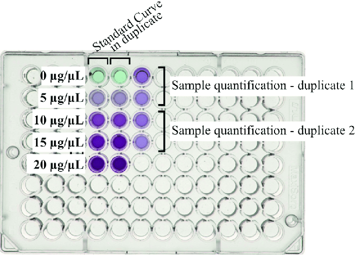
- Sample Preparation: Begin by preparing the protein sample, ensuring it is appropriately diluted and free from contaminants that could interfere with the quantification process.
- Assay Setup: Follow the instructions provided with the protein quantification kit to set up the assay. Add the sample and the reagents required for the specific assay, and mix thoroughly.
- Incubation: Incubate the assay mixture for the specified duration to allow the color to develop, which is indicative of the protein concentration in the sample.
- Measurement: Use a spectrophotometer or a plate reader to measure the absorbance of the samples at the appropriate wavelength. Compare the absorbance values to a standard curve generated using known concentrations of a protein standard.
- Calculation: Utilize the standard curve to calculate the protein concentration in the sample. Take into account any dilutions made during the sample preparation process.
- Quality Control: Perform quality control checks to ensure the accuracy and reproducibility of the results. Repeat the assay if necessary to validate the findings.
- Data Interpretation: Analyze the results and interpret the data to draw conclusions about the protein concentration in the original sample.
Protein Electrophoresis
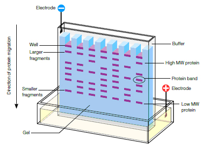
Protein electrophoresis is a vital technique used to separate and analyze proteins based on their size and charge. To begin, prepare protein samples using appropriate buffers and denaturing agents like SDS. Subsequently, cast polyacrylamide gels of the necessary concentration to facilitate effective protein separation. Load the prepared samples onto the gel wells and apply an electric current for migration. Once the proteins have separated, utilize suitable staining methods like Coomassie Brilliant Blue or silver stain for visualization. Finally, destain the gel and analyze the separated protein bands, comparing them to known molecular weight markers to determine protein characteristics and concentrations.
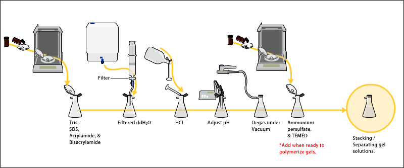
PAGE Gel Preparation
To prepare a polyacrylamide gel electrophoresis (PAGE) gel, start by creating a resolving gel and a stacking gel with the desired acrylamide concentration. Allow the gels to polymerize, and then carefully remove the comb to create sample wells. Next, equilibrate the gel with the running buffer and load the protein samples into the wells. Finally, run the gel at the appropriate voltage for the necessary separation time, monitoring the progress using appropriate staining techniques or protein visualization methods.
During the preparation of a polyacrylamide gel electrophoresis (PAGE) gel, the resolution of proteins is achieved by their differential migration through a polyacrylamide matrix, which separates proteins based on their size and charge. The PAGE gel is used in various techniques, including SDS-PAGE, native PAGE, and two-dimensional gel electrophoresis, providing insights into protein structure, purity, and molecular weight determination.
Marker
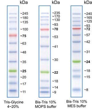
Protein markers are essential tools in gel electrophoresis techniques, aiding in the determination of molecular weight and the evaluation of gel performance. They are utilized by mixing with loading buffer and heating to denature before being loaded onto the gel. Subsequently, the gel is either stained directly or transferred to a membrane for Western blot analysis or other detection methods, allowing for accurate assessment and analysis of protein samples during the electrophoresis process.
To use protein markers, they are typically mixed with loading buffer and denatured by heating before being loaded onto the gel. After electrophoresis, the gel is stained or transferred to a membrane for Western blot analysis or other detection methods. Protein markers are used to estimate the molecular weight of protein samples and to monitor the progress and efficiency of electrophoresis experiments. They serve as reference points to compare the migration of target proteins and assess the overall success of the separation process.
Fast Blocking
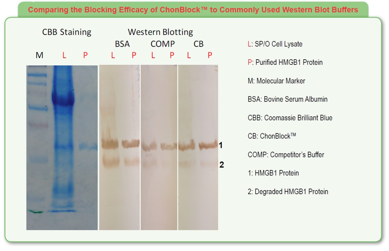
Gentaur offers two types of blocking reagents: BioTeZ Blocking Solution and ChonBlock.
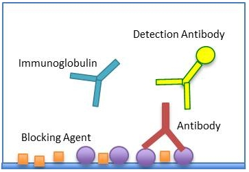
BioTeZ Blocking Solution is a protein-based blocking reagent composed of BSA. It is a universal blocking reagent that can be used for a variety of assays, including ELISA, Western blotting, and immunohistochemistry. BioTeZ Blocking Solution is effective at blocking non-specific binding and is compatible with a wide range of antibodies.
ChonBlock is a non-protein-based blocking reagent composed of a proprietary blend of polymers. It is a high-performance blocking reagent that is specifically designed for ELISA assays. ChonBlock is highly effective at blocking non-specific binding and does not interfere with the assay signal.
Here is a table summarizing the key features of each blocking reagent:
| Feature | BioTeZ Blocking Solution | ChonBlock |
|---|---|---|
| Type | Protein-based | Non-protein-based |
| Composition | BSA | Proprietary blend of polymers |
| Applications | ELISA, Western blotting, immunohistochemistry | ELISA |
| Effectiveness | Effective at blocking non-specific binding | Highly effective at blocking non-specific binding |
| Compatibility | Compatible with a wide range of antibodies | Does not interfere with the assay signal |
The choice of blocking reagent depends on the specific requirements of the assay. BioTeZ Blocking Solution is a good general-purpose blocking reagent, while ChonBlock is a high-performance blocking reagent that is specifically designed for ELISA assays.
Blocking Reagent
A blocking reagent is a substance used in various biochemical assays to prevent non-specific binding and minimize background signal interference. It is typically added to reaction mixtures to occupy unbound sites on the surface of the membrane or other materials, preventing subsequent binding of detection reagents and reducing false-positive signals. This process is crucial in immunological techniques like Western blotting and ELISA, ensuring accurate and reliable results by enhancing the specificity and sensitivity of the assay.
Protein Electrophoresis
Protein electrophoresis is a commonly employed technique in biochemistry and molecular biology to separate and analyze proteins based on their size and charge. It involves the migration of proteins through a gel matrix under the influence of an electric field, with their movement influenced by factors such as size, shape, and charge. This process allows for the characterization and quantification of proteins, enabling researchers to study their structure, function, and interactions in various biological contexts.
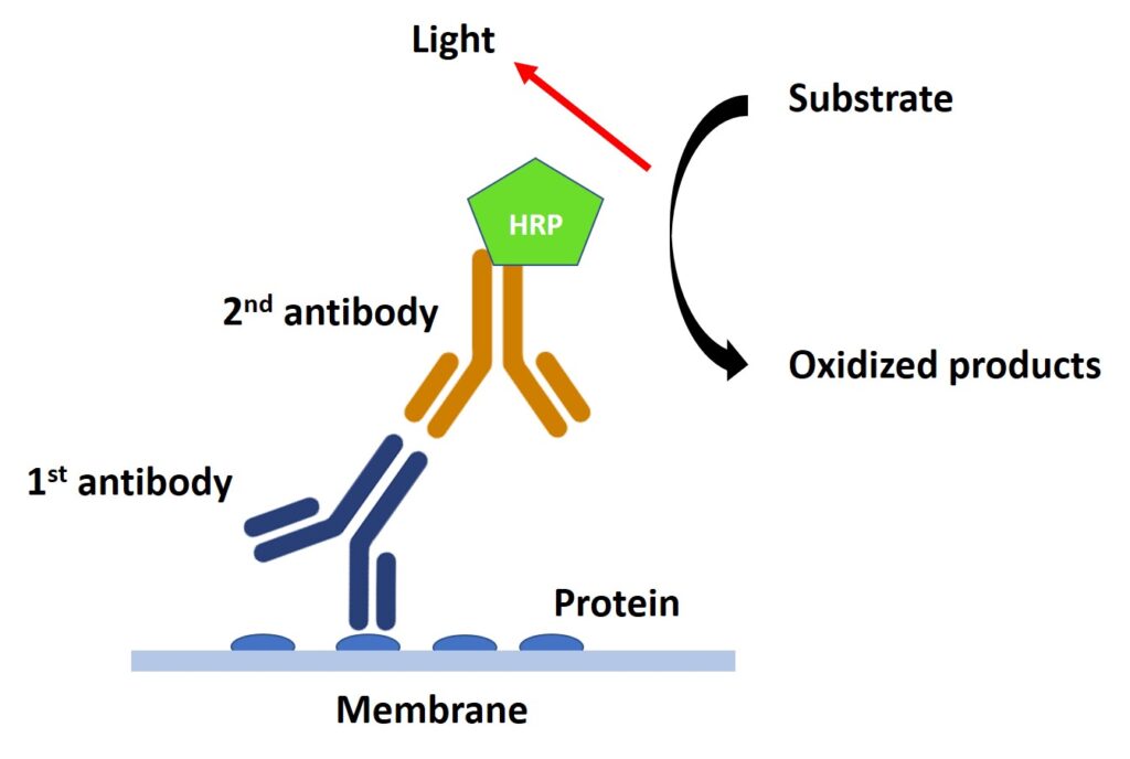
ECL
Enhanced ECL Chemiluminescent Substrate Kit is a high-performance chemiluminescent substrate designed to detect proteins labeled with horseradish peroxidase (HRP) in Western blot and ELISA applications. It offers enhanced sensitivity, prolonged signal duration, and reduced background compared to conventional ECL substrates.
Key Features:
- High Sensitivity: Enables detection of proteins at picogram levels.
- Prolonged Signal Duration: Optical signals last up to 5 hours, allowing for multiple exposures.
- Reduced Background: Minimizes background interference, enhancing signal clarity.
- Easy to Use: Convenient and requires no special optimization of operating steps.
Applications:
- Western Blotting: Detect proteins of interest in Western blots.
- ELISA: Quantify proteins in ELISA assays.
Kit Components:
- Substrate A: Contains the chemiluminescent substrate.
- Substrate B: Contains the stabilizing reagent.
- Enhancer: Enhances signal intensity and duration.
Storage:
- Store all components at 2-8°C.
Advantages:
- Enhanced sensitivity for low-abundance protein detection.
- Prolonged signal duration for multiple exposures and image analysis.
- Reduced background for improved signal clarity.
- Easy-to-use protocol for seamless integration into existing workflows.
Overall, the Enhanced ECL Chemiluminescent Substrate Kit from Gentaur is a valuable tool for researchers seeking high-sensitivity, low-background detection of HRP-labeled proteins in Western blotting and ELISA applications.
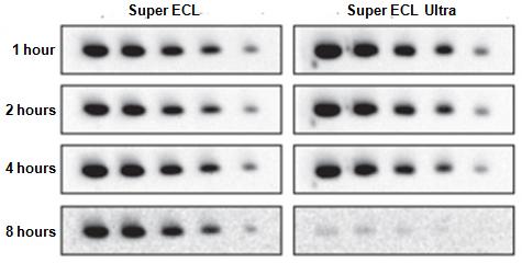
Super ECL Detection Reagent ECL
Super ECL Detection Reagent is a highly sensitive and effective chemiluminescent substrate used in Western blotting and other protein detection techniques. It enables the detection of low levels of target proteins, offering enhanced sensitivity compared to standard ECL reagents. With its optimized formulation, the Super ECL Detection Reagent provides rapid and efficient visualization of specific proteins, making it a valuable tool in various molecular biology applications.

SuperSignal SuperDura Extended Duration Substrate
The SuperSignal SuperDura Extended Duration Substrate is an advanced chemiluminescent detection reagent optimized for enhanced sensitivity and signal longevity in various Western blotting and protein detection applications. Offering prolonged signal duration, this substrate is particularly effective for visualizing low-abundance target proteins and ensuring precise quantification in complex biological samples. With its long-lasting and stable chemiluminescent signal, the SuperSignal SuperDura Extended Duration Substrate enables accurate and reliable protein analysis, contributing to the success of diverse research studies in the field of molecular biology.

SuperSignal MaxiSignal Maximum Sensitivity Substrate
The SuperSignal MaxiSignal Maximum Sensitivity Substrate is an advanced chemiluminescent detection reagent that offers unparalleled sensitivity in Western blotting and protein detection applications. With its exceptional ability to detect even trace amounts of proteins, this substrate is specifically designed to amplify signal intensity, allowing for the precise visualization and analysis of low-abundance target proteins. The SuperSignal MaxiSignal Maximum Sensitivity Substrate is an invaluable tool for researchers aiming to achieve optimal sensitivity and accurate quantification in their molecular biology experiments, thereby enhancing the reliability and quality of their research outcomes.
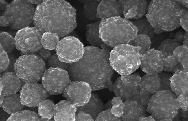
Affinity Gel
Affinity gel is typically used in various biochemistry and molecular biology techniques for the purification of proteins, peptides, or nucleic acids. To utilize it, first, equilibrate the gel with an appropriate buffer and then add the sample containing the target molecule. Allow sufficient time for the target molecule to bind to the ligand on the gel. Next, wash the gel to remove any nonspecifically bound impurities. Finally, elute the target molecule using an elution buffer suitable for the specific affinity interaction. When working with affinity gel, it is crucial to handle it with care to prevent any contamination and maintain the integrity of the bound target molecule. Proper storage conditions should also be followed to preserve the functionality and effectiveness of the affinity gel.
Protein G Affinity Gel is a high-performance affinity chromatography resin designed for the purification of monoclonal and polyclonal antibodies from various sources, including serum, ascites fluid, and cell culture supernatants. This affinity gel utilizes Protein G, a bacterial protein with a high binding affinity for the Fc region of IgG antibodies.
Key Features:
- High Binding Capacity: Efficiently captures IgG antibodies from various sources.
- High Specificity: Binds selectively to IgG antibodies, minimizing non-specific binding.
- Ease of Use: Convenient and straightforward purification protocol.
Applications:
- Antibody Purification: Purify monoclonal and polyclonal antibodies from various sources.
- Immunoglobulin G (IgG) Isolation: Isolate IgG from complex biological samples.
Kit Components:
- Protein G Affinity Gel: Pre-packed columns or bulk resin.
- Buffers: Binding buffer, washing buffer, and elution buffer.
Storage:
- Store the resin at 2-8°C. Store the buffers according to their respective instructions.
Advantages:
- High binding capacity for efficient antibody capture.
- High specificity for IgG antibodies, minimizing contamination.
- Easy-to-use protocol for rapid antibody purification.
Overall, Protein G Affinity Gel from Gentaur is a valuable tool for researchers seeking a reliable and efficient method for purifying antibodies from various sources.
Immunity Magnetic Beads
Magnetic Beads
Magnetic beads are widely used in various molecular biology and biochemical applications for the isolation and purification of biomolecules such as nucleic acids, proteins, and cells. These beads typically have a functionalized surface that specifically binds to the target biomolecule. The binding occurs under appropriate buffer conditions and is facilitated by a magnetic field, allowing for the separation of the beads from the solution. After isolation, the target biomolecule can be eluted from the beads using an elution buffer suited for the specific interaction. Care should be taken to ensure proper handling of magnetic beads to avoid contamination and to maintain the integrity of the isolated biomolecules.
Nucleic Acid Removal
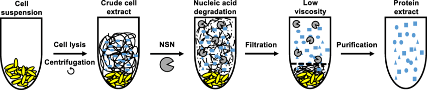
Nucleic acid removal involves the elimination of unwanted DNA and RNA from biological samples or solutions. This process is crucial in various applications, such as protein purification, to avoid interference or contamination from nucleic acids. Common methods for nucleic acid removal include enzymatic digestion, affinity chromatography, and magnetic bead-based techniques. Care should be taken to choose a method that effectively removes nucleic acids while preserving the integrity and functionality of the target protein. Additionally, the choice of the appropriate protocol depends on the specific characteristics of the sample and the downstream application.
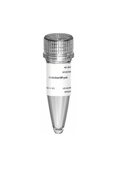
UltraNuclease
For price : Contact us
UltraNuclease refers to a highly efficient enzyme used for the specific degradation of nucleic acids, including both DNA and RNA molecules. This enzyme is often employed in various laboratory procedures such as the elimination of contaminating nucleic acids during protein purification, reducing non-specific interactions in molecular biology assays, and the prevention of carryover contamination in PCR reactions. UltraNuclease is known for its robust activity across a broad range of temperatures and pH conditions, making it a versatile tool for nucleic acid removal in various research and biotechnological applications.
Gentaur UltraNuclease is a highly active and versatile enzyme product designed to eliminate DNA and RNA from protein samples. It is a mixture of four high-performance nucleases that effectively degrade a wide range of nucleic acids, including single-stranded DNA (ssDNA), double-stranded DNA (dsDNA), single-stranded RNA (ssRNA), and double-stranded RNA (dsRNA).
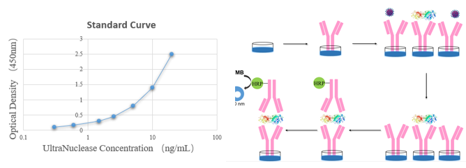
UltraNuclease Detection
UltraNuclease Detection involves the identification and quantification of the activity of UltraNuclease enzymes within a biological or experimental sample. This detection process often utilizes specific assays or techniques that can measure the enzymatic degradation of nucleic acids in the presence of UltraNuclease. These assays are crucial for confirming the efficacy and activity of UltraNuclease in removing or degrading unwanted nucleic acids, ensuring the integrity and purity of biological samples for downstream applications.
- Store all components at -20°C.
- High sensitivity for detecting low levels of UltraNuclease activity.
- High specificity for accurate detection without interference.
- Quantitative measurement for assessing enzyme activity levels.
- Ease of use for convenient application in various settings.